High-Plex Spatial Profiling
of Suspension Cells
SmartBioSurface® enables reliable high-plex spatial profiling of suspension cells, supporting integrated analysis of blood-derived and tissue samples within a unified workflow.
When combined with advanced cyclic immunofluorescence platforms such as CellScape™, CellScape XR™, and Quanterix Spatial PhenoCycler®, SmartBioSurface® provides the stable cell immobilization required for repeated staining, washing, and imaging cycles while preserving image quality and assay reproducibility.
Exceptional cell retention across multiple imaging cycles ensures consistent data generation and minimizes sample loss. In cyclic immunofluorescence workflows, SmartBioSurface® achieved approximately 98.5% cell retention after ten imaging cycles, supporting robust and reproducible biomarker analysis. Combined with the multiplexing capabilities of CellScape XR™, this enables high-content profiling with excellent signal quality.
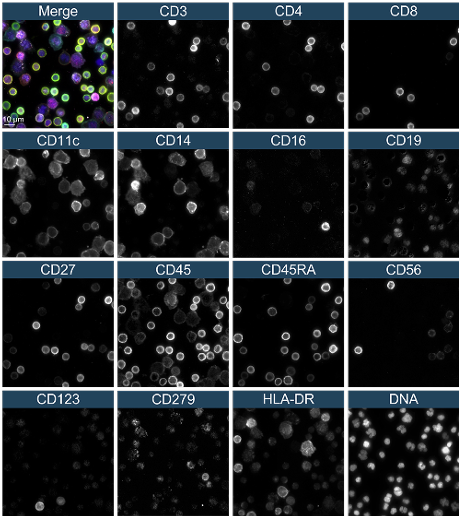
SmartBioSurface® has also demonstrated compatibility with the Quanterix Spatial PhenoCycler® platform, maintaining cell integrity throughout repeated staining and reporter-exchange procedures. Multiplex analysis of peripheral blood leukocytes generated specific and reproducible staining patterns across multiple immune markers, confirming the suitability of the workflow for high-dimensional immune profiling.
By enabling the use of validated reagent panels and imaging chemistries across different platforms and sample types, SmartBioSurface® facilitates direct comparison between tissue sections and suspension-derived cells. This integrated approach provides a more comprehensive view of complex biological systems and supports advanced applications in immuno-oncology, immunology, infectious diseases, and translational research.
Scalable, reproducible, and platform-compatible, SmartBioSurface® provides a robust foundation for next-generation spatial biology studies of non-adherent cells.